
FACT SHEET: Oral Cancer (includes “oral cavity cancer” and “oropharyngeal cancer”, sometimes referred to as “oral and oropharyngeal cancer” [OC/OPC])1
Is the initiation of non-invasive dental hygiene procedures* contra-indicated?
- Possibly (dental hygiene procedures should not be scheduled while the patient/client is experiencing oral ulcerations and pain, has an acute oral infection, has an absolute neutrophil count ≤ 1.0 X 10⁹/L, or has a platelet count ≤ 50 X 109/L)
Is medical consult advised?
- Possibly (e.g., if suspicious lesion is detected; if intraoral infection and/or immunosuppression is suspected, particularly if the patient/client is undergoing radiation therapy and/or chemotherapy)
Is the initiation of invasive dental hygiene procedures contra-indicated?**
- Possibly (contra-indicated for persons undergoing radiotherapy and/or chemotherapy for oral cancer); furthermore, dental hygiene procedures should not be scheduled while the patient/client is experiencing oral ulcerations and pain, has an acute oral infection, has an absolute neutrophil count ≤ 1.0 X 10⁹/L, or has a platelet count
- ≤ 50 X 109/L
Is medical consult advised?
- See above.
Is medical clearance required?
- Yes, if the patient/client is about to undergo or is undergoing active chemotherapy or radiation therapy for oral cancer.
- Yes, if the patient/client is scheduled for major oral surgery for oral cancer.
Is antibiotic prophylaxis required?
- No, not typically (although cancer or treatment-induced immunosuppression may warrant consideration of antibiotic prophylaxis).
Is postponing treatment advised?
- Possibly (depends on whether cancer and its treatment may interfere with invasive procedures and whether there is immunosuppression associated with cancer treatment).2
Oral management implications
- Dental hygienists play an important role in early detection of oral cancer, leading to timely medical/dental referral and potential biopsy, endoscopy, and imaging. Patient/client survival is much better when the cancerous lesion is diagnosed at an early stage. 25% of oral cancers occur in non-smokers who have no other known significant risk factors.
- The dental hygienist should determine risk factors by reviewing the patient/client’s medical history, with specific attention to past history of oral cancer, specific risk-related habits, and reasons for hospitalizations. Two of the most important modifiable risk factors for oral cancer are tobacco and alcohol use, which account for up to 75% of cases. Tobacco- and alcohol-associated lesions tend to occur on the anterior tongue and in the anterior mouth.
- Increased risk of cancer holds for all types and uses of tobacco, whether it is smoked as a cigarette, cigar, pipe, bidi (a small, hand-rolled cigarette commonly used in Asia) or hookah, or used smokeless as plug, chew, or snuff. Stopping tobacco consumption reduces the risk of oral cancer and premalignant lesions, although it may take 10 to 20 years for a former smoker’s risk to reduce to that of a nonsmoker.
- “Reverse smoking”, the habit of holding the lit end of a cigarette inside the mouth (as is a habit in India and some South American countries) is associated with a particularly high risk of oral cancer.
- Exposure to second-hand smoke may increase the risk of oral cancer in non-smokers.
- Tobacco and alcohol consumption work together synergistically, greatly increasing risk of oral cancer. Heavy smokers and drinkers are also more likely to be diagnosed with late-stage disease. Questions about tobacco and alcohol use should include frequency (current and past use), and amount and duration of use; responses should be recorded and updated regularly.
- Patients/clients from South Asian countries (e.g., India, Pakistan, Bangladesh, and Sri Lanka) may chew betel quid, which is a carcinogenic mixture of plant components that often contains tobacco. Dental hygienists should enquire about past and current consumption of betel quid, areca nut, or paan, and cessation should be advised.
- Smoking cannabis increases the risk of oral cancer. (Marijuana smoke contains many of the same carcinogens found in tobacco smoke and has four times the tar burden.)
- In recent years in North America, human papilloma virus (HPV) — particularly types HPV-16 and HPV-18 — has been increasingly implicated in oral cancer. HPV-related oropharyngeal cancers are usually found in the lingual and palatine tonsils, the soft palate, and the base of the tongue. Most patients/clients with HPV-related cancers of the head and neck are 50 to 60 years of age, about 10 years younger than typical patients with oropharyngeal cancer. Incidence is also rising in adults younger than 50 years old. Risk factors include higher number of sexual partners, higher number of oral sex partners, and early age at first intercourse.
- In contrast to smoking-related head and neck cancers, survival outcomes for HPV-related cancers are very good. Such cancers are usually treated with radiation and concurrent chemotherapy; however, such treatment can lead to swallowing dysfunction and feeding-tube dependence.
- HPV immunization greatly reduces rates of oropharyngeal infection, and this will likely translate into reduced incidence of HPV-related oropharyngeal cancers in the future. In order to prevent HPV-related oral cancer, dental hygienists can play a role in suggesting that unvaccinated patients/clients — both male and female, particularly those aged 9 to 26 years — speak with their primary care physician regarding possible immunization. Currently in Ontario, HPV vaccine is publicly funded for all female and male Grade 7 students.
- Immunosuppression (due to medications, disease, or bone-marrow transplantation) is a risk factor for oral cancer.
- Sun exposure is a well-established risk factor for lip cancer. Dental hygienists should promote the use of sun blocking lip balms with their patients/clients.
- A diet rich in fruits and vegetables reduces the risk of premalignant lesions and oral cancer. Chronic irritation in the mouth may increase risk.
- Chronic psoriasis and lichen planus of the mouth elevate risk. Persons of Ashkenazi Jewish ancestry are at higher than average risk, as are persons with a family history of squamous cell carcinoma of the head and neck.
- Inspection and palpation of cervical neck nodes (including submandibular and submental nodes) by the dental hygienist is important, because oral cavity cancer may metastasize to the lymphatic system. In addition to intraoral examination, inspection and palpation of the salivary glands, lips, and cheeks is also important for the detection of oral cancer.
- Before the initiation of chemotherapy and radiotherapy, oral evaluation and treatment should be scheduled. If all nonsurgical dental and dental hygiene procedures have not been accomplished before initiation of head or neck radiotherapy, they should be performed within the first 2 weeks of therapy before the onset of mucositis.
- During oral radiation therapy, the radiation oncologist should direct perioral tissue care. Some lip balms and lubricants can potentiate the effects of local radiation and result in significant radiation dermatitis. Furthermore, patients/clients with dentures should be advised to leave the dentures out of their mouths as often as possible to reduce mucositis.
- Salivary gland dysfunction or xerostomia associated with radiotherapy or chemotherapy can be managed by usual methods. The dental hygienist should ensure that the patient/client about to undergo radiation therapy is assessed by a physician or dentist for consideration of pilocarpine hydrochloride; this medication can decrease the severity of radiation-induced xerostomia.
- Oral infections associated with chemotherapy can be reduced by frequent oral hydration and optimal bacterial plaque control. Use of antifungals that are sugar-free should be encouraged. The dental hygienist should ensure medical consult occurs (i.e., oncologist is alerted) at first signs of oral infection. Early detection and treatment of oral infections are important to prevent exacerbation of mucositis that may require cancer therapy interruption.
- Bleeding related to chemotherapy-induced myelosuppression is not preventable, but consistent removal of bacterial plaque can minimize this complication. Referral to an oncologist should occur if low platelet count is suspected on history taking (e.g., tendency to easily bleed/bruise; presence of petechiae [pinpoint hemorrhages on skin]) or if excessive bleeding occurs during oral procedures.
- Rampant dental caries or tooth demineralization may result from cancer therapy-induced salivary gland dysfunction. Prevention and management measures include bacterial plaque control, frequent oral hydration, daily application of 1.1% sodium fluoride gel in custom gel containers (designed to extend beyond cervical line of the teeth) or topical fluoride, in-office application of fluoride varnish to exposed cementum, and dietary counseling to discourage snacking on cariogenic and acidic foods/drinks. If dental decay occurs despite daily fluoride application, the patient/client can be placed on a 2-week chlorhexidine regimen and in-office fluoride varnish application.
- Trismus or temporomandibular disorder related to the direct effect of radiotherapy may be present and the dental hygienist should ensure referral to an appropriate health care professional for management. Dental hygiene procedures may need to be altered for patients/clients with trismus to avoid exacerbation of pain (e.g., shortened appointments or sedation).
- All teeth within the field of radiation that have a poor lifetime prognosis should be extracted 14 to 21 days prior to the initiation of radiotherapy. Surgical insult should be avoided to irradiated bone throughout the patient/client’s lifetime. Referral to an oral surgeon should occur for surgical (and possible hyperbaric oxygen) management of necrotic tissue and bone. Frequent and regular dental hygiene care is indicated to reduce periodontal disease.
Oral manifestations
- Oral cancer includes cancer of the lips, cheeks, gums and teeth, floor of the mouth, tongue, oropharynx, tonsils, and salivary glands.3
- The most common sites of oral cancer are:
- lateral border of the tongue (30%)
- floor of the mouth (14%)
- soft palate complex (posterior soft palate, uvula, and faucial arches) (11%)
- lower lip (38%)4.
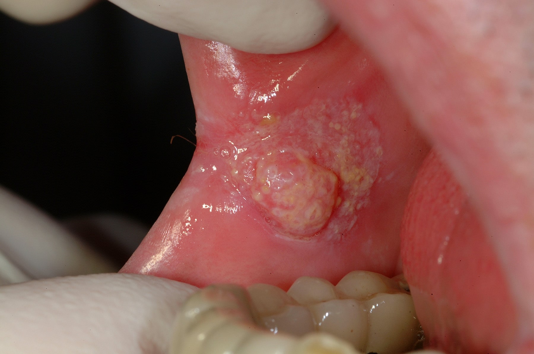
- Oral squamous cell carcinoma (SCC) accounts for 90% to 95% of all cancers in the oral cavity. Oral SCC is variable in appearance; it often manifests as a painless swelling or exophytic ulcerative mass, but early tumours may present as leukoplakia or erythroplakia. Recurrences after treatment are frequent, especially if patients continue to use tobacco and alcohol.
- Symptoms/signs of oral SCC include hoarseness, dysphagia, dysarthria, lumps, intractable ulcers, bleeding, numbness, pain, loosening of teeth, difficulty opening mouth, change in fit of dentures, and, in advanced cancer, difficulty in breathing and loss of gag reflex (from nerve involvement that may result from soft palate involvement).
- SCC may occur at elevated rates in patients/clients with long-standing oral lichen planus (LP), particularly LP of an erosive or atrophic nature.
- Other head and neck tumours include basal cell carcinoma (which does not occur in the oral cavity, but rather on the skin or lips, and which presents as a nonhealing ulcer with characteristic rolled borders); salivary gland tumours (major and minor glands; intraorally, minor gland tumours are most commonly located at the junction of the hard and soft palates, but may also occur on the labial — usually the upper lip — and buccal mucosa, the retromolar area, the floor of the mouth, and rarely the tongue); odontogenic tumours derived from tooth-forming tissues (only rarely malignant, but can be locally destructive to mandible and/or maxilla); uncommon tumours of nerves, muscles, blood vessels, and melanin-producing cells (including malignant melanoma, which usually presents as a rapidly enlarging blue-to-black mass, most typically intraorally on the palate and maxillary gingiva; dental hygienists should remember the ABCDEs — Asymmetry, Border, Colour, Diameter, Evolving —of melanoma detection; although primary malignant melanoma of the oral cavity is rare, lesions arising on the skin may metastasize to the mouth); bone and cartilage; blood-forming tissues (such as lymphoma, for which the most common intraoral location is the tonsillar area; dental hygienists should also be alert to swellings of lymphoid tissue at the base of the tongue and soft palate, as well as swelling of lymph nodes); and metastatic tumours of the jaws (which usually arise from primary cancers of the thyroid, breast, prostate, lungs, and kidneys, with the most frequent intraoral site being the mandible; signs and symptoms include pain, paresthesia/anesthesia of the lip, swelling, expansion of affected bone, and loosening of teeth in involved area).
- Unlike most traumatic or infective lesions, cancer does not show healing within two weeks. Instead, the cancerous lesion shows change in colour, shape, and size over time. Common signs of oral cancer are chronicity (failure to heal, particularly within 14 days); erythroplakia (red patch that is smooth, granular, or velvety that cannot be diagnosed as any other disease without biopsy); fixation (immobility); induration (hardness); leukoplakia (white, plaque-like lesion that cannot be wiped off and cannot be diagnosed as any other disease without biopsy); lymphadenopathy (firmness and enlargement of lymph nodes and tonsils); and ulceration (loss of epithelial integrity).
- There is a higher risk of cancerous transformation in erythroplakia than in leukoplakia, although both are associated with oral cancer. However, leukoplakia is 60 times more common than erythroplakia.
- Late-stage treatment for oral cancer usually involves major orofacial surgery, radiation, and chemotherapy.
- Treatment for oral cancer may result in mucositis and/or stomatitis (from direct effect of radiation therapy and cytotoxic chemotherapy); salivary gland dysfunction or xerostomia (from direct radiation damage to salivary gland tissue and possible indirect effect of chemotherapeutic agents); infection (related to chemotherapy-induced immunosuppression — fungal, viral, and bacterial); bleeding (related to chemotherapy-induced myelosuppression); trismus (limited ability to open the mouth) or temporomandibular disorder (due to direct effect of radiation on muscles of mastication and/or TMJ); and soft tissue necrosis and osteoradionecrosis (related to direct effect of radiation on tissue and bone).
- Salivary gland dysfunction is usually permanent after radiation therapy, whereas function usually returns after chemotherapy.
- Oral infections in patients/clients undergoing chemotherapy may not cause typical signs and symptoms. Candidiasis is common during radiation therapy.
- Alteration in taste commonly occurs during radiation therapy to the tongue and mouth. Damage to the taste buds may partially or completely resolve after completion of radiotherapy.
- Dysphagia is a potential acute complication of radiation to the head and neck.
- Telangiectasia and friable mucosa may be chronic sequelae of radiation therapy, as are altered tooth and jaw development in children.
- Trismus usually occurs 3 months after high dose radiotherapy to the masticatory muscles or TMJ ligaments and remains a lifelong problem. In addition to discomfort, it can interfere with eating, talking, and post-treatment examination.
- Surgery for oral cancer has the following potential complications: infection; airway obstruction; fistula formation; necrosis in the surgical site; impairment of swallowing and speech, smell, hearing and vision; drooling; malocclusion; temporomandibular disorders; and facial deformity.
Related signs and symptoms
- More than 75% of head and neck cancers originate in the oral cavity. In 2020, oral cancer was newly diagnosed in approximately 5,400 residents of Canada, and it was responsible for about 1,500 deaths. Like most cancers, incidence of oral cancer rises with age and risk factor duration, with patients/clients over 60 years of age being at greatest risk. However, the incidence of oral cancer is increasing in persons younger than 40 years of age, likely due to changing risk factors (e.g., HPV infection).
- Overall, men are twice as likely to be afflicted as women. However, the ratio is almost 1:1 in patients/clients under 40. With growing immigration from high-risk areas such as India (where oral cancer accounts for up to 50% of all malignancies), the number of cases of oral cancer may continue to increase in Canada.
- About 1 in 67 Canadian men is expected to develop oral cancer during his lifetime, and 1 in 200 will die from it.
- About 1 in 142 Canadian women is expected to develop oral cancer during her lifetime, and 1 in 333 will die from it.
- Persistent earache in only one ear, numbness or tingling in the mouth or face, persistent or recurrent cough/sore throat/upper respiratory tract infection, and feeling like there is a lump or something caught in the throat can be symptoms/signs of oral cavity or oropharyngeal cancer.
- In addition to the oropharynx and oral cavity, HPV-related cancers also occur in the throat, cervix, vagina, and anus, as well as the genitalia of both sexes.
- In addition to lesions in the oral cavity, smoking-related cancers may also present with patients/clients complaining of hoarseness, persistent cough, weight loss, and blood in the urine.
- Oral squamous cell carcinoma spreads by local infiltration into adjacent tissues or metastasis to regional lymph nodes through lymphatic channels. Spread to local structures causes induration, fixation, and lymphadenopathy. Carcinomas of the floor of the mouth, tongue, and posterior sites tend to metastasize earlier than lesions located in anterior oral sites such as the lips. Lesions in the maxillary region have a greater tendency to metastasize than those in the mandibular region. While distant metastasis is rare, the most common sites for dissemination are the lungs, liver, and bone. This cancer often results in death, with the overall 5-year survival being about 50%.
- Acute potential complications from radiotherapy to the head and neck include impaired nutrition (from xerostomia, pain, and dysphagia), hearing loss, and fatigue.
- Complications from head and neck surgery may include impairment of hearing, vision, and smell; compromised nutritional status; and chronic pain in the shoulder muscles. There may also be profound psychosocial problems related to facial disfiguration and/or guilt regarding high-risk habits (e.g., smoking).
References and sources of more detailed information
- College of Dental Hygienists of Ontario
https://cdho.org/advisories/oral-cancer/
https://cdho.org/advisories/gastrointestinal-tract-tumours/ - Tranby EP, Heaton LJ, Tomar SL, Kelly AL, Fager GL, Backley M, Frantsve-Hawley J. Oral Cancer Prevalence, Mortality, and Costs in Medicaid and Commercial Insurance Claims Data. Cancer Epidemiol Biomarkers Prev. 2022 Sep 2;31(9):1849-1857. doi: 10.1158/1055-9965.EPI-22-0114. PMID: 35732291; PMCID: PMC9437560.
https://www.ncbi.nlm.nih.gov/pmc/articles/PMC9437560/# - Rivera C. Essentials of oral cancer. Int J Clin Exp Pathol. 2015 Sep 1;8(9):11884-94. PMID: 26617944; PMCID: PMC4637760.
https://www.ncbi.nlm.nih.gov/pmc/articles/PMC4637760/ - Laronde DM, Hislop TG, Elwood JM and Rosin MP. Oral Cancer: Just the Facts. J Can Dent Assoc 2008;74:3.
www.cda-adc.ca/jcda/vol-74/issue-3/269.html - Palma DA, Nichols AC. Human papillomavirus in head and neck cancer. CMAJ. 2014;186(5):370. doi:10.1503/cmaj.130849
https://www.ncbi.nlm.nih.gov/pmc/articles/PMC3956567/ - Poh CF, Williams PM, Zhang L, Rosin MP. Heads Up! – A Call for Dentists to Screen for Oral Cancer. J Can Dent Assoc. 2006;72(5):413-416.
https://www.cda-adc.ca/jcda/vol-72/issue-5/413.pdf - Public Health Ontario
https://www.publichealthontario.ca/-/media/documents/hpv-vaccine-technical.pdf?la=en - Government of Canada
https://www.canada.ca/en/public-health/services/oral-diseases-conditions/oral-cancer.html
https://www.canada.ca/en/public-health/services/reports-publications/health-promotion-chronic-disease-prevention-canada-research-policy-practice/vol-35-no-1-2015/supplement/page-27.html - Dental Hygiene Canada
https://www.dentalhygienecanada.ca/oralcancer - Canadian Cancer Society
www.cancer.ca/en/cancer-information/cancer-type/oral
https://cancer.ca/en/research/cancer-statistics/canadian-cancer-statistics
- Canadian Dental Association
http://www.cda-adc.ca/en/oral_health/talk/complications/diseases/oral_cancer.asp - Ontario Dental Hygienists Association
https://odha.on.ca/wp-content/uploads/2016/08/Oral-Cancer-Screening.14.1-copyright.pdf - Ontario Dental Association
https://www.oda.ca/oral-health-basics/oral-conditions-diseases/oral-cancer/ - Medline Plus, U.S. National Library of Medicine, National Institutes of Health
https://medlineplus.gov/ency/article/001035.htm (Oral Cancer) - Dentistry IQ
https://www.dentistryiq.com/dentistry/oral-cancer/article/14299226/the-dental-hygienists-guide-to-oral-pathology-in-patients-with-oral-cancer - Today’s RDH
https://www.todaysrdh.com/how-hygienists-can-be-the-first-line-of-defense-against-oral-cancer/ - Medscape
https://emedicine.medscape.com/article/2047890-overview (Oral Cavity Cancer Treatment Protocols) - Bowen DM (ed.) and Pieren JA (ed.). Darby and Walsh Dental Hygiene: Theory and Practice (5th edition). St. Louis: Elsevier; 2020.
- Little JW, Miller CS and Rhodus NL. Little and Falace’s Dental Management of the Medically Compromised Patient (9th edition). St. Louis: Elsevier; 2018.
- Ibsen OAC and Phelan JA. Oral Pathology For The Dental Hygienist (6th edition). St. Louis: Saunders Elsevier; 2014.
- Regezi JA, Sciubba JJ and Jordan RCK. Oral Pathology: Clinical Pathologic Correlations (7th edition). St. Louis: Elsevier; 2017.
FOOTNOTES
1 Increasingly, the term “oral cancer” is used to encompass both oral cavity cancer and oropharyngeal cancer. Some authorities, however, reserve the term “oral cancer” for oral cavity cancer.
2 When a suspicious lesion is newly detected by the dental hygienist, invasive procedures should be implemented cautiously or even avoided altogether in the area of concern to prevent trauma to or alteration of the lesion prior to the dental hygienist’s obtaining medical advice.
3 Oral cavity cancer sites are sometimes defined as including the inner lip, parts of the tongue apart from the base and lingual tonsil, gingiva, floor of the mouth, palate, and “other unspecified parts of the mouth.” Oropharyngeal cancer, according to some definitions, occurs at sites including the base of the tongue, the lingual tonsil and tonsil, the oropharynx, and the pharynx including Waldeyer’s ring. The most-common sites of HPV-related head and neck squamous cell carcinoma (HNSCC) are the tonsils and base of tongue within the oropharynx; HPV-related HNSCC is uncommon in nonoropharyngeal sites.
4 Carcinomas of the lower lip are much more common than upper lip lesions, although the prognosis for upper lip lesions is considerably worse. In addition to smoking (especially pipe smoking), ultraviolet light (i.e., sun) exposure is a significant risk factor for carcinoma of the lips. Lip carcinomas appear most commonly in patients/clients between 50 and 70 years of age and affect men much more frequently than women.
* Includes oral hygiene instruction, fitting a mouth guard, taking an impression, etc.
** Ontario Regulation 501/07 made under the Dental Hygiene Act, 1991. Invasive dental hygiene procedures are scaling teeth and root planing, including curetting surrounding tissue.